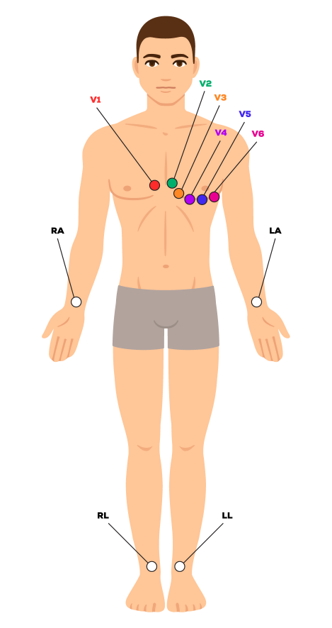
12-Lead ECG Placement Guide

What is ECG Lead Placement?
ECG Lead Placement is the process of attaching electrodes to the patient’s body in order to measure and record the electrical activity of the heart. EKG readings can also detect various heart conditions and previous heart attacks.
Correct lead placement can help ensure accurate ECG readings and provide valuable insight into a patient’s heart health. Depending on the type of ECG, the number of leads and their placements can vary.
Understanding the Basics of Lead Placement
Correct lead placement allows medical professionals to accurately measure the electrical activity of the heart and read and analyze EKG results. Each lead typically measures one particular type of electrical activity for a specific location on the patient’s body. The placement of the leads should be consistent across all ECG’s and can be adjusted as needed to ensure accurate readings.
Standard 12-lead ECG Leads
The standard ECG, often referred to as 12-lead ECG, includes 12 leads obtained with 10 electrodes. An electrode is a conductive pad that is attached to the skin and enables electrical current recording.
The 12-lead ECG consists of four limb leads (RA, RL, LA, LL) and six precordial (chest) leads (V1, V2, V3, V4, V5, and V6).
A Guide to 12-Lead ECG Placement
Placing ECG leads can be a challenging process, but with a few simple steps, it can be easy to ensure accurate lead placement. Here is a step-by-step guide to correctly placing ECG leads:
Preparing the Patient
Before placing the ECG leads, it is important to ensure that the patient is adequately prepared. This includes make sure that the patient’s skin is clean and dry, and that any clothing is removed or loosened to allow for proper lead placement.
Preparing the patient also includes…
- Ensuring patient is in a supine position with arms at their sides
- Shaving hair that can interfere with electrodes
- Removing clothes that can interfere with electrodes
- Confirming electrode gel is moist
- and more
Placing the Leads
In a 12-lead ECG, there are 10 electrodes that provide various angles on the heart’s activity.
For standard ECG, place the limb leads on the patient’s arms and legs, and place the precordial leads on the patient’s chest.
Chest Electrodes and Placement
- V1 – Fourth intercostal space on the right sternum
- V2 – Fourth intercostal space at the left sternum
- V3 – Midway between placement of V2 and V4
- V4 – Fifth intercostal space at the midclavicular line
- V5 – Anterior axillary line on the same horizontal level as V4
- V6 – Mid-axillary line on the same horizontal level as V4 and V5
Limb Electrodes and Placement
- RA (Right Arm) – Anywhere between the right shoulder and right elbow
- RL (Right Leg) – Anywhere below the right torso and above the right ankle
- LA(Left Arm) – Anywhere between the left shoulder and the left elbow
- LL (Left Leg) – Anywhere below the left torso and above the left ankle


