Complete Guide to Ultrasound Probes

What is an Ultrasound Probe?
Ultrasound probes, also known as ultrasound transducers, allow ultrasound machines to capture real-time images of a patient’s anatomy. When it comes to ultrasound imaging, choosing the right probe is crucial. With various probe types available, selecting the right probe can significantly impact the quality of your imaging. Whether you are scanning abdominal organs, vascular structures, or performing cardiac assessments, understanding the differences between linear, convex, phased array, and other transducer types is key. In this article, we’ll guide you through the main types of ultrasound probes, breaking down their specific uses and helping you determine which one is best for your application.
Types of Ultrasound Probes
Convex Probe
A convex ultrasound transducer, also referred to as a curvlinear transducer, produces a wide field of view at a lower frequency. Convex transducers emit ultrasound waves in a fan-shaped beam, making them useful for imaging structures within the body such as internal organs and deep tissues.
Key Features
- Curved shape: The probe surface is convex, which allows for a larger scanning area and a wider field of view.
- Lower frequency: Convex probes operate at lower frequencies (2-5 MHz), making them suitable for deeper penetration. However, the image resolution is lower.
- Common uses: Convex transducers are widely used for abdominal, obstetric, and gynecological imaging. Convex transducers are able to visualize larger and deeper areas in the body, such as the uterus, liver, kidneys, and fetal structures during pregnancy.
Linear Probe
A linear ultrasound transducer is a type of ultrasound probe that produces sound waves in a straight line. These transducers generate high-frequency sound waves, resulting in detailed, high-resolution images for small areas.
Key Features
- Flat shape: The transducer’s surface is flat, producing a rectangular field of view.
- High frequency: Operates at higher frequencies (5-15MHz), providing excellent resolution for imaging structures that are closer to the skin surface.
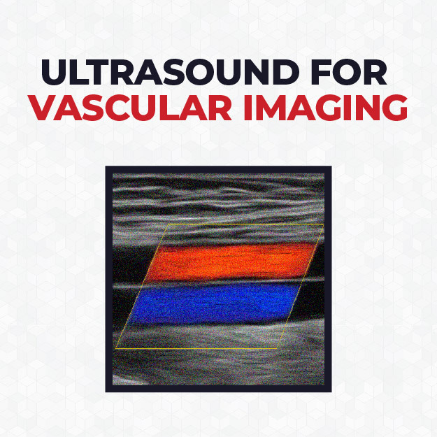
- Common uses: Linear transducers are typically used for vascular imaging, musculoskeletal scans, nerve, small parts and more.
Phased Array Probe
A phased array transducer is a type of ultrasound probe that uses a small array to produce sound waves at different angles. Unlike other types of transducers, all the elements in a phased array transducer fire almost simultaneously but with slight time delays, allowing the beam to be steered electronically without moving the probe.
Key Features
- Beam steering: Sound waves are emitted at various angles by electronically controlling the timing of the pulses. This allows for creating a fan-like image that can visualize a wide area from a narrow probe position.
- Lower frequency: Phased array transducers operate at lower frequencies (1-5 MHz), allowing for deeper penetration but lower resolution compared to higher-frequency probes.
- Common uses: Phased array transducers excel at imaging deep organs and areas that require a wide field of view despite limited access. These transducers are primarily used for cardiac imaging , abdominal imaging, and transcranial applications.
Endocavity Probe
An endocavity transducer is designed to enter a body cavity, such as the rectum or vagina, to obtain detailed images of internal organs. Endocavity transducers provide clear, close-up images of internal organs and tissues that are difficult to visualize with external ultrasound methods.
Key Features
- High frequency: Endocavity probes typically operate at higher frequencies (5-9 MHz), providing detailed, high-resolution images with less depth penetration.
- Probe shape: The transducer has a long, slender design with a curved or linear array at the tip to facilitate insertion into body cavities.
- Common Uses: For detailed imaging of the uterus, ovaries, and surrounding pelvic structures, often used in gynecology and obstetrics.
Transesophageal Echo Probe
A Transesophageal Echo (TEE) Transducer is a critical tool in cardiac imaging, providing high-resolution images of the heart’s structures and blood flow without interference from the lungs, ribs, or other tissues. TEE transducers are especially useful in diagnosing heart conditions that are difficult to assess with traditional, non-invasive ultrasound techniques.
Key Features
- Flexible, Long Probe Design: The TEE probe’s design allows it to be passed down the patient’s throat into the esophagus.
- High-Quality Heart Imaging: TEE probes provide superior images of the heart’s chambers, valves, and large blood vessels.
- Common Uses: These transducers are used to guide cardiac surgeries, such as valve repairs or replacements.